

Dynamic Liquid Surface Enhanced Raman Scattering Platform Based on Soft Tubular Microfluidics for Label-Free Cell Detection
Introduction:
Surface-enhanced Raman spectroscopy (SERS) refers to the phenomenon where enhanced electromagnetic fields on sample surfaces significantly enhance the Raman scattering signals compared to ordinary Raman scattering (NRS) signals when certain molecules are adsorbed onto rough metal surfaces. It has been proven to be a quick and efficient spectroscopic method. SERS has high detection sensitivity and is easily applicable for detecting weak signal samples, but in practical applications, the repeatability and detection limits of SERS technology have always been challenging. Therefore, the repeatability and sensitivity of SERS technology have always been a focus of scientific research.
Research findings:
Recently, Hu Bo’s group from the School of Life Sciences at Xi’an University of Electronics Technology proposed a dynamic fluid SERS platform based on hose microfluidics, effectively addressing current issues and successfully applying it to the detection and classification of breast cancer cells. It holds great promise in cell research, clinical diagnosis, and food safety.
Comparison of advantages and disadvantages between traditional testing methods and microfluidic technology:
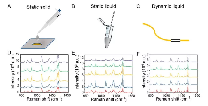
The traditional SERS measurement methods include static solid-phase and static liquid-phase approaches. Static solid-phase SERS detection often fails to obtain uniform spectral signals with uniform intensity due to the difficulty in achieving uniform mixing and distribution between analytes and nanoparticles on a substrate. While static liquid phase can address the uniform dispersion of analytes and nanoparticles at spatial scales to some extent, it is difficult to ensure spectral reproducibility due to uncertain mixing times, irregular scattering, local heating, and photodegradation. Moreover, both methods also require preprocessing steps such as pre-mixing and drying, as well as long mixing times.
Microfluidic technology, when used in conjunction with SERS technology to form a microfluidic-SERS system, enables continuous acquisition of SERS spectra under dynamic flow conditions, thereby enhancing the repeatability of SERS detection while increasing detection efficiency.
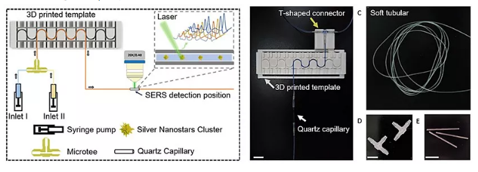
However, traditional microfluidic chip fabrication requires expensive clean rooms and the process is time-consuming and labor-intensive. Other microfluidic chip processing methods that do not require cleanrooms still require expensive and specialized instruments, limiting their processing accuracy to some extent. The team from Xi’an Electronic Science and Technology University uses a flexible microfluidic SERS platform embedded with commercial, biocompatible, and transparent Microsoft tubes in 3D printing support templates, enabling dynamic solution mixing, precise control of mixing time, and continuous SERS spectrum acquisition.
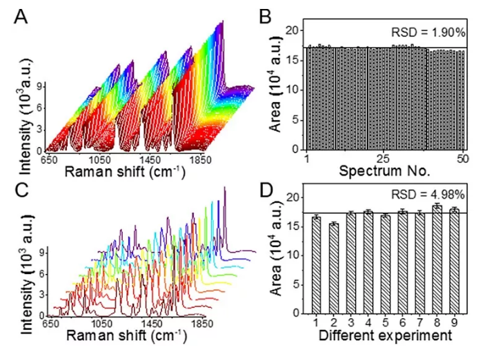
Compared to traditional static solid-phase and static liquid-phase measurement methods, the dynamic fluid SERS platform demonstrates good stability and repeatability by characterizing model molecules with relative standard deviations of 1.90% and 4.98%, respectively.
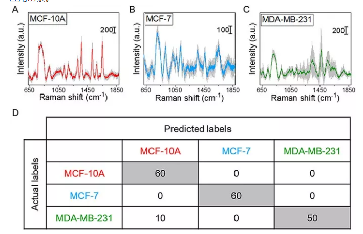
In addition, the platform was used to collect Raman spectra of three types of breast cells: normal (MCF-)10A breast cells and mild malignant (MCF-7) breast cancer cells, combined with classification and identification using the K-NN algorithm, achieving sensitivity over 83.3%, specificity over 91.6%, and accuracy of 94.4%.
The dynamic fluid SERS platform based on hose microfluidic control offers advantages such as low processing costs and simple manufacturing methods, enriching the processing and preparation technology of microfluidic chips. Moreover, it can collect a large number of continuous fluids’ SERS spectra in a short amount of time, offering advantages such as speed, versatility, sensitivity, reliability, low cost, and no need for preprocessing. The platform is poised to become a powerful tool for detecting unlabeled cells in liquid environments and holds broad application prospects in cellular research, clinical diagnosis, and food safety.
This achievement was recently published in Analytical Chemistry and was completed by Hu Bo’s research group at Xi’an University of Electronic Science and Technology. The first authors of the article are Xuan Xiaoding, a master’s student, and Zhao Lei, a postdoc. link Https://pubs.acs.org/doi/abs/10.1021/acs.analchem.9b01111
This study employed the “Finder One” micro-area laser Raman spectroscopy system from Beijing Zhuoli Hangguang Instrument Co., Ltd. For more information about this product, please feel free to consult our company.
Disclaimer
The content (including images) posted on the official account of Beijing Zhuoli Hangguang Instrument Co., Ltd. comes from sources provided by the original author or reprinted with permission. The article’s copyright, data, and stated views belong to the original author. Beijing Zhuoli Hangguang Instrument Co., Ltd. The purpose of publishing and reprinting is to convey more information and facilitate online sharing.
If you believe this article contains any infringement, please contact us so that we can address it promptly. We strive for rigorous and accurate data, and we look forward to your valuable insights if you have any questions. We also warmly welcome your submissions and feedback.