0
EVIDENT (Olympus) cellSens 이미징 소프트웨어 Imaging Software
Life science microscopy imaging software with 5D acquisition, TruAI deep learning, deconvolution, and live-cell analysis
5D 획득, TruAI 딥러닝, 디컨볼루션, 생세포 분석 지원 생명과학 이미징 소프트웨어
Description
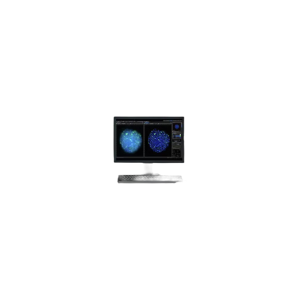
cellSens™ is Evident’s comprehensive life science microscopy imaging software platform, designed to streamline image acquisition, processing, and analysis workflows. With an intuitive customizable interface and powerful multi-dimensional imaging capabilities, cellSens supports researchers from routine documentation to complex live-cell experiments and AI-assisted analysis.
cellSens™는 Evident의 생명과학 현미경 이미징 소프트웨어 플랫폼으로, 이미지 획득·처리·분석 워크플로를 간소화합니다. 직관적이고 커스터마이즈 가능한 인터페이스와 강력한 다차원 이미징 기능을 갖추어, 일상적 문서화부터 복잡한 생세포 실험 및 AI 기반 분석까지 지원합니다.
Key Features
- Customizable user interface with pre-configured layouts for various research workflows
- 다양한 연구 워크플로를 위한 사전 구성 레이아웃 및 커스터마이즈 가능한 UI
- 5D image acquisition (X, Y, Z, T, wavelength) with Graphical Experiment Manager (GEM)
- 그래픽 실험 관리자(GEM)를 통한 5차원 이미지 획득 (X, Y, Z, T, 파장)
- TruSight™ deconvolution including GPU-accelerated 3D Constrained Iterative algorithm
- GPU 가속 3D Constrained Iterative 알고리즘을 포함한 TruSight™ 디컨볼루션
- TruAI™ deep learning for automated image segmentation and cell/nucleus detection
- 자동 이미지 분할 및 세포/핵 검출을 위한 TruAI™ 딥러닝
- Real-time panoramic stitching, EFI, and HDR imaging
- 실시간 파노라마 스티칭, EFI(확장 초점 이미징), HDR 이미징
- Well Plate Navigator for automated multi-well screening
- 자동화된 멀티 웰 스크리닝을 위한 웰 플레이트 탐색기
- Object tracking, count & measure, FRET/FRAP analysis tools
- 객체 추적, 계수 및 측정, FRET/FRAP 분석 도구
- Conference mode for real-time collaboration and remote microscopy
- 실시간 협업 및 원격 현미경 검사를 위한 컨퍼런스 모드
Applications
- Live-cell imaging: cell migration, division, and long-term time-lapse studies
- 생세포 이미징: 세포 이동, 분열 및 장기 타임랩스 연구
- Fluorescence multi-channel imaging and spectral unmixing
- 형광 다채널 이미징 및 스펙트럼 언믹싱
- High-content screening and drug discovery using well plate formats
- 웰 플레이트 형식을 이용한 고함량 스크리닝 및 신약 개발
- Neuroscience: brain slice and organoid imaging
- 신경과학: 뇌 절편 및 오가노이드 이미징
- Cell biology: confluency measurement, cell counting, tracking
- 세포생물학: 컨플루언시 측정, 세포 계수, 추적
- Cancer research and stem cell studies with 3D imaging
- 3D 이미징을 이용한 암 연구 및 줄기세포 연구
Specifications
| Feature | Entry | Standard | Dimension |
|---|---|---|---|
| Image Acquisition | Snapshot/Video | + Time-lapse, Multi-wavelength | Full 5D (XYZT + λ) |
| EFI / Panoramic MIA | Option | Manual Process | ● |
| TruSight Deconvolution | – | – | ● (CI Deconv. option) |
| TruAI Deep Learning | – | Option | Option |
| Well Plate Navigator | – | – | Option |
| Count & Measure | – | Option | Option |
| FRET/FRAP Analysis | – | – | Option |
| OS | Windows 10 Pro (22H2) / Windows 11 Pro (24H2) 64-bit | ||
| Recommended RAM | 16 GB+ (32 GB+ for deep learning) | ||
| Compatible Cameras | Evident DP/SC/LC/UC series, Hamamatsu ORCA series, Andor, Photometrics, and more | ||
| Supported File Formats | Read/Write: JPEG, TIFF, PNG, BMP, AVI, OIR, VSI, PSD, Big TIFF; Read: OIF/OIB, STK, MRC | ||